Successful Installation of First SPECS Momentum Microscopy System
The first KREIOS MM analyzer was recently installed at the UVSOR Synchrotron in Okazaki, Japan
Momentum Microscopy is an emergent application in the field of photoemission spectroscopy. It is especially useful for researchers who are mostly working with topological insulators, 2D materials such as Graphene, or who focus on Fermi Surface mapping of their materials under investigation. Another exciting future trend in this area is the acquisition of spin-resolved Momentum Microscopy images.
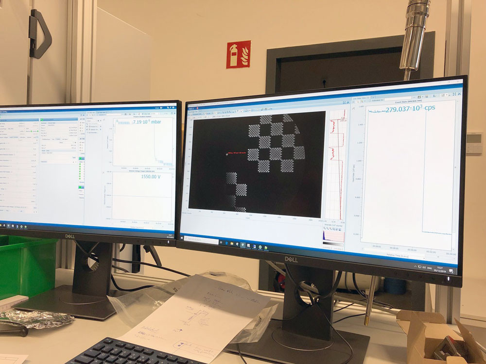
The KREIOS and KREIOS MM combine a hemispherical analyzer with a momentum microscopy column as its lens to image the complete band structure of a sample surface in a single measurement. Real space (PEEM) images of the sample surface can also be recorded with a resolution better than 100 nm / 50 nm respectively, in order to select an analysis region of interest and perform spectroscopy on selected small areas down to <2 um. These features are unheard of in classical ARPES analyzers.
The small spot analysis capability of the KREIOS 150 MM calls for the highest possible flux density of the photon source employed for photoelectron excitation. It is therefore a perfect product to be installed at synchrotron beamlines, making use of the high brilliance and focus. For laboratory applications advances are also being made with improved manufacturing techniques and newly designed optics. The UV source with a new generation of specially designed optics boosts the photoncflux density by a factor of >10 compared to established light sources such as the UVS 300 ETC.
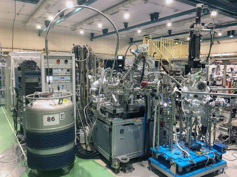
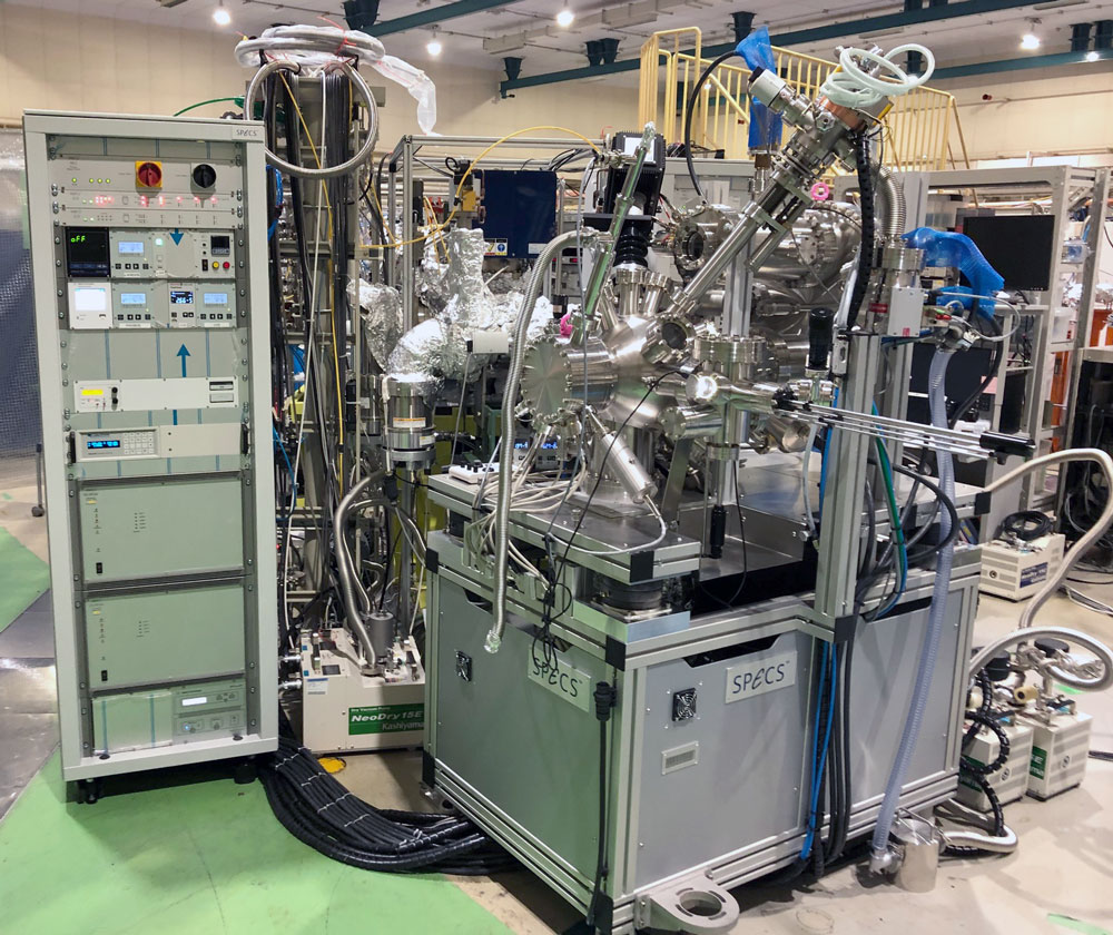
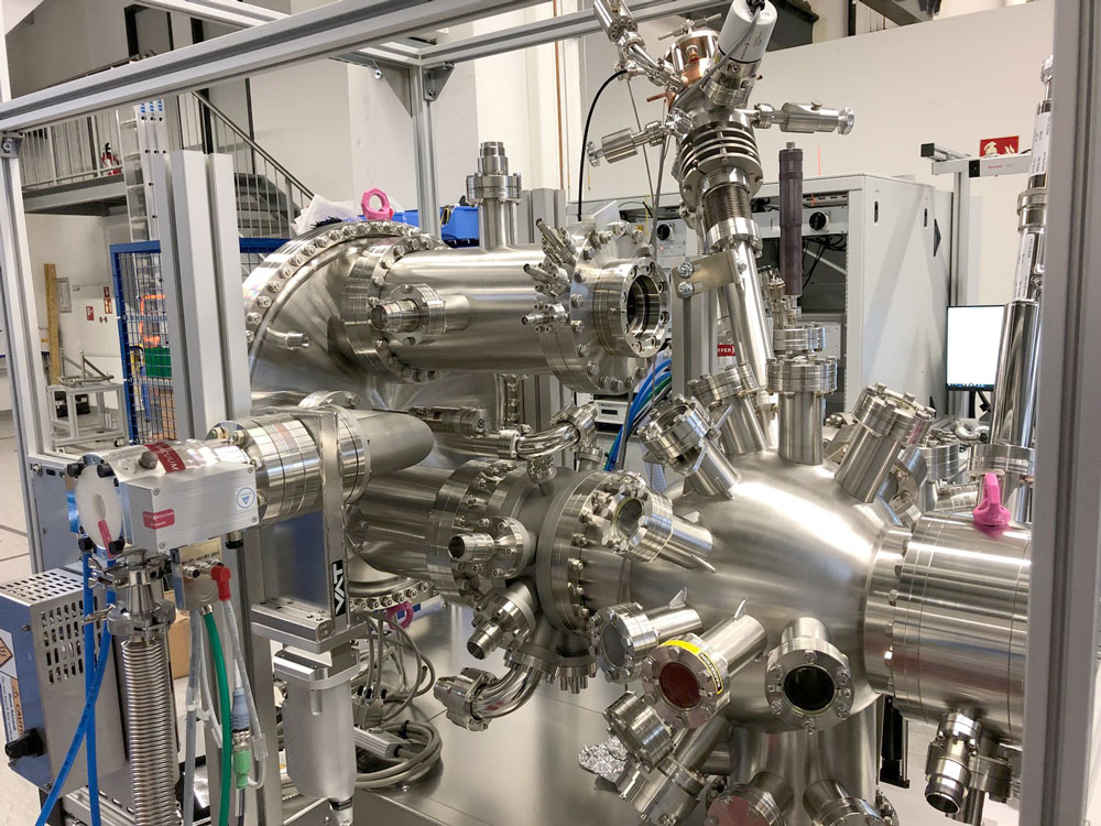
SPECS are proud to announce the successful installation of the first KREIOS MM system at the Japanese UVSOR synchrotron facility located in Okazaki. The system was installed at the end station and will be taken into commission immediately by the responsible scientists.
An upgrade of this already powerful combined ARPES/PEEM/MM system, adding a second hemisphere and an imaging spin filter, which projects the full momentum microscopy image across a spin-filtering crystal, has already been confirmed and are scheduled successively for the coming two years.